As Plastic surgeons, we are frequently involved in the reconstruction of defects after excision of jaw tumors. Hence, it’s good to have a fair idea about the types of tumors of the mandible and their classifications. We have earlier posted the various classifications for Mandibular defects that are usually created after excision of these tumors.
Residents note: It’s a commonly asked question in exams in India and its details are not available in popular Plastic Surgery books but in Dental books.
Classification (💡SIMPLIFIED)
This jaw tumor classification is based on the tissue of origin, histology and whether they are benign or malignant and is adapted from the WHO classification of odontogenic and maxillofacial bone tumors (2017), a part of WHO classification of head and neck tumours (4th edition).
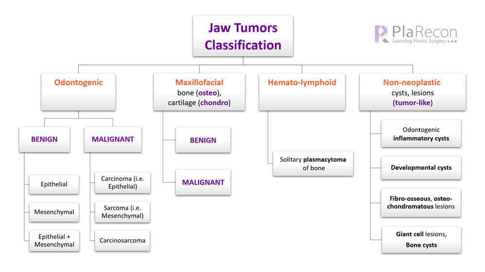
- Odontogenic (💡TEETH + GUMS)
- Benign
- Epithelial
- Mesenchymal
- Epithelial + Mesenchymal (MIXED)
- Malignant
- Carcinoma (i.e. Epithelial)
- Sarcoma (i.e. Mesenchymal)
- Carcinosarcoma (MIXED)
- Benign
- Maxillofacial Bone and Cartilage (💡FACIAL BONES-osteo, CARTILAGES-chondro)
- Benign
- Malignant
- Hematolymphoid (💡HEMATOPOIETIC, LYMPHOID TISSUES)
- Non-neoplastic cysts and lesions (💡TUMOR-LIKE)
- Odontogenic inflammatory cysts
- Odontogenic and non-odontogenic developmental cysts
- Fibro-osseous and osteochondromatous lesions
- Giant cell lesions and bone cysts
Note: Odontogenic cysts though not ‘tumors’ i.e. neoplastic, are conceptually inseparable from the odontogenic tumors (which may also be cystic)- and hence have been included in the 2017 WHO Classification of Odontogenic tumors, cysts and allied lesions. Moreover, with more genetic studies of the tumor and a better understanding of the etiopathogenesis and behaviour of the tumors, some entities like Keratocystic odontogenic tumor (WHO, 2005) have now been shifted to cyst category as odontogenic keratocyst (WHO, 2017).
Classification (in detail)
Benign odontogenic tumours
Epithelial
- Ameloblastoma: unicystic/ extraosseous or peripheral/ metastasising types
- Squamous odontogenic tumour
- Calcifying epithelial odontogenic tumour (Pindborg tumor)
- Adenomatoid odontogenic tumour
💡Mnemonic: ASCA
Mesenchymal
- Odontogenic fibroma
- Odontogenic myxoma/myxofibroma
- Cementoblastoma
- Cemento-ossifying fibroma
Epithelial and Mesenchymal
- Ameloblastic fibroma
- Primordial odontogenic tumour
- Odontoma
- Dentinogenic ghost cell tumour
Malignant odontogenic tumours
Odontogenic carcinomas
- Ameloblastic carcinoma
- Primary intraosseous carcinoma, not otherwise specified
- Sclerosing odontogenic carcinoma
- Clear cell odontogenic carcinoma
- Ghost cell odontogenic carcinoma
Odontogenic sarcomas
Odontogenic carcinosarcoma
Benign maxillofacial bone and cartilage tumours
- Chondroma
- Osteoma
- Melanotic neuroectodermal tumour of infancy
- Chondroblastoma
- Chondromyxoid fibroma
- Osteoid osteoma
- Osteoblastoma
- Desmoplastic fibroma
Malignant maxillofacial bone and cartilage tumours
- Chondrosarcoma
- Osteosarcoma
Hematolymphoid tumours
- Solitary plasmacytoma of bone
Non-neoplastic Cysts and Lesions
As mentioned above, these cysts or (tumor-like) lesions that may present as swelling in the jaw region and technically are not tumor or neoplasm.
- Odontogenic inflammatory cysts
- Radicular cyst*
- Inflammatory collateral cysts
- Odontogenic and non-odontogenic developmental cysts
- Dentigerous cyst*
- Odontogenic keratocyst
- Lateral periodontal cyst and Botryoid odontogenic cyst
- Gingival cysts
- Glandular odontogenic cyst
- Calcifying odontogenic cyst
- Orthokeratinized odontogenic cyst
- Nasopalatine duct cyst
- Fibro-osseous and osteochondromatous lesions
- Ossifying fibroma
- Familial gigantiform cementoma
- Fibrous dysplasia
- Cemento-osseous dysplasia
- Osteochondroma (Exostosis)*
- Giant cell lesions and bone cysts
- Giant cell granuloma- central, peripheral
- Cherubism* (Familial multilocular cystic disease of the jaws)
- Aneurysmal bone cyst
- Simple bone cyst
With so many classifications of jaw tumors— Kramer, Pindborg, Shear histological classification (1992), WHO (2005), modified WHO (2017)— this topic becomes confusing at times.
💡 So simply remembering the headings as mentioned in the Simplified version of the classification (at the top) and then remembering a few examples of each by following the histologic or tissue of origin patterns and few starred* ones may aid in remembering this long classification.

Tutorials & tips in Plastic Surgery
+ Weekly updates of high-quality webinars!
