The most characteristic feature of the human hand is the comparatively ‘longer and opposable thumb’. Thenar muscles are part of the intrinsic muscles of the hand that contributes to the finer movements of the thumb including the opposition.
Level– Basics
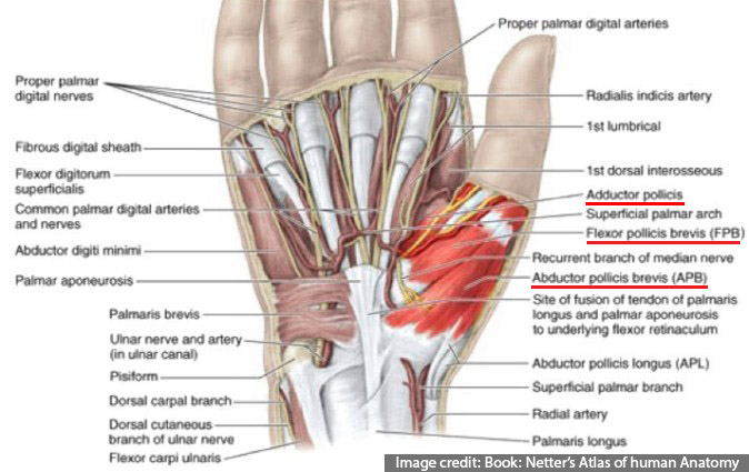
Thenar Eminence
The Thenar eminence comprise of a group of the following four* muscles, which are supplied mainly by the Median nerve:
- Abductor pollicis brevis (APB)
- Opponens pollicis (OPP)
- Flexor pollicis brevis (FPB)
- *Adductor pollicis (ADD) – 💡 supplied by the Ulnar nerve and hence, is not considered by some as a part of thenar muscles.1 Note: Ulnar nerve also supplies the deep head of the FPB.
The origin of most of the thenar muscles is partly from the transverse carpal ligament (flexor retinaculum)
Their insertions are primarily on the base of the proximal phalanx of thumb, except for the opponens, which inserts on the first metacarpal.
Regardless of the innervation, thenar muscles are supplied by C8 and T1 roots.
Blood Supply: Superficial palmar branches of Radial artery, which arises from the radial artery at the level of the radial styloid.

Opposition of the Thumb
A thumb in true opposition is not only opposite the fingers, but it is far forward from them and is rotated, so the pulp faces the fingers and nail parallel the palm. Opposition is a rotational movement of the first metacarpal around the long axis of its shaft. The motion of opposition “consists of the rotation of the volar surface of the thumb from the 90o of rest to 180o– parallel with the transverse plane of the hand, so that it is able to face the volar surface of the fingers after pulling out completely (abduction) from the surface of the palm.”
Opposition involves the combined motions of flexion, pronation, and palmar abduction of the thumb metacarpal. Reposition is the opposite of opposition.
Other movements of the Thumb
Abduction is moving the thumb anterior to, or away from, the palm. Adduction is bringing the thumb metacarpal toward the second metacarpal, in the plane of the palm. Flexion is moving the thumb in an ulnar direction within the plane of the palm. Extension is the opposite motion of flexion.
Thenar muscles individually:
1. Abductor pollicis brevis (APB)
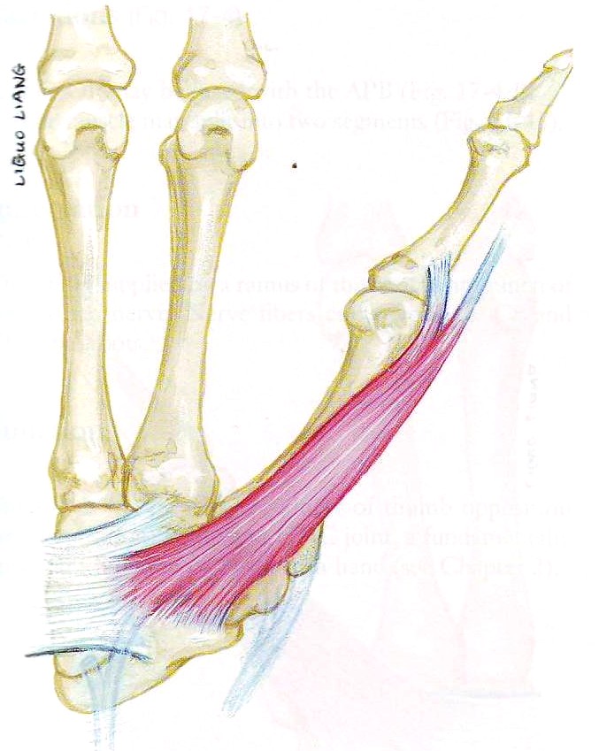
Most important thenar muscle. Superficial, flat, thin muscle- provides the bulk to the thenar eminence.
Origin: Flexor retinaculum, tubercle of Scaphoid,+/- tubercle of Trapezium. Insertion: Lateral side of base of PPx of thumb + capsule of MCP joint.
Actions: Abduction and flexion of the metacarpal (MC), slight flexion of the proximal phalanx, extension of the distal phalanx (through its EPL insertion). Pronation of the entire thumb occurring through the Carpo-MC (CMC) joint is simultaneous with flexion of the thumb MC. These actions produce opposition. These actions are enhanced by OPP.
2. Opponens pollicis (OPP)
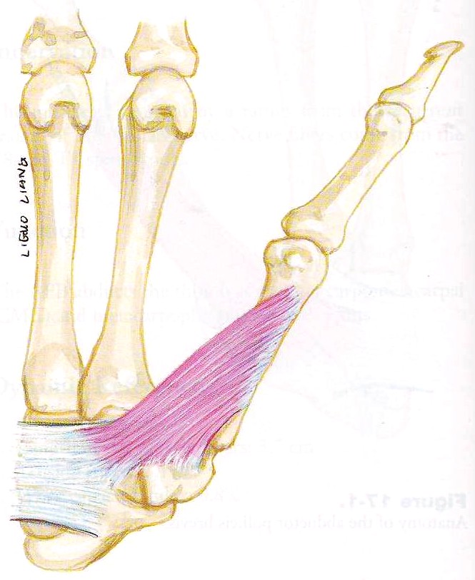
Short, thick, triangular muscle. Located below APB.
Origin: Flexor retinaculum, tubercle of Trapezium. Insertion: Whole length of the MC of thumb – radial side.
Arterial supply: Superficial palmar branch of the radial artery + branches from the first palmar meta- carpal artery, princeps pollicis, radialis indicis arteries; and the deep palmar arch.
Action: Initiation of opposition– Flexion, pronation of thumb MC at CMC jt. Enhances work of opposition of APB.
3. Flexor pollicis brevis (FPB)
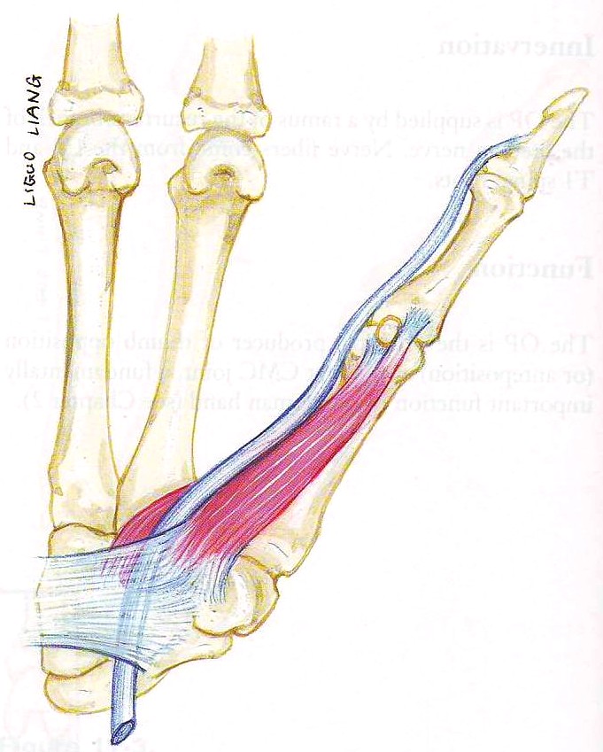
Superficial part: Origin: Flexor retinaculum, tubercle of Trapezium. Insertion: Radial side of base of PPx of thumb (Note: tendon has a sesamoid bone)
Deep part: Origin: Ulnar side of the first metacarpal bone. Insertion: ulnar side of base of PPx of thumb along with AP.
Arterial supply: Superficial palmar branch of the radial artery + branches from princeps pollicis and radialis indicis arteries.
Action: Flexion at MCP jt. Additional actions include extension of the distal phalanx and pronation of the thumb MC.
4. Adductor pollicis (ADD)
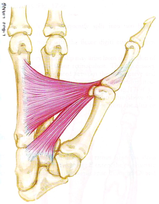
Flat, fan-shaped, triangular, deepest.
Oblique head– Origin: Capitate bone, Bases of 2nd and 3rd MC, intercarpal ligaments, tendon sheath of FCR. Insertion: ulnar side of base of PPx of thumb along with FPB, tendon has a sesamoid bone.
Transverse head– Origin: Lower 2/3rd- palmar surface- 3rd MC. Insertion: Ulnar side of base of PPx of thumb along with FPB.
Arterial supply: The princeps pollicis, radialis indicis arteries (occasionally combined as the first palmer metacarpal artery), and branches from the deep palmar arch.
Action: Primary action is adduction of the thumb metacarpal. The muscle also extends the thumb interphalangeal joint via its insertion onto the dorsal apparatus of the thumb.
Note: The deep palmar arch and the deep motor branch of the ulnar nerve pass between the two heads of ADD. The first dorsal interosseous muscle lays on the dorsum of the ADD and, together, these two muscles provide the mass of the first web space. Volarly, ADD is crossed by the index finger flexor tendons and the first lumbrical.
Variations:
Riche-Cannieu anastomosis
![Riche-Cannieu anastomosis [CB- Connecting Branch]](https://www.plarecon.com/wp-content/uploads/2015/12/Riche-Cannieu-anastomosis.jpg)
Neural connections from the Deep palmar branch of the Ulnar N. to the recurrent branch of Median N. in the hand (ulnar-to-median in hand). Hence, fibres from the Ulnar N. may supply the OPP and/or APB (which are normally innervated by the median nerve- “Exception” in hand- along with the 1st and 2nd lumbricals).
Note: There are many other communications between the Median and Ulnar nerves in the hand and forearm.
Bibliography
- Atlas of Human Anatomy- by Netter.
- Atlas of Hand Anatomy and Clinical Implications- by Han-Liang Yu, Robert Chase, and Berish Strauch.
- Nadire Unver Dogan et al. The communications between the ulnar and median nerves in upper limb. Neuroanatomy 2009-8: 15–19
- Gupta, Jost. Anatomy and Function of the Thenar Muscles. Hand Clin 28 (2012) 1–7

Tutorials & tips in Plastic Surgery
+ Weekly updates of high-quality webinars!
